On the 7th and 8th of May, the International ME/CFS Conference 2026 took place in Berlin and online, and ME Research UK was in attendance remotely.
Talks covered a wide range of topics relating to ME/CFS, and to long COVID, including genetics, autoimmunity, neuroinflammation, endothelial dysfunction. Although generally inconclusive, the results of the latest research into potential treatments were also discussed, such as hyperbaric oxygen therapy, low-dose naltrexone, methylprednisolone, low-dose rapamycin, and daratumumab.
As with previous years, sessions at the 2026 conference were recorded, and will be available to watch online in due course.
Highlights of the conference included a presentation on the results from the DecodeME study and discussion of next steps, and a presentation on whole-body positron emission tomography (PET) scans for ME/CFS.
Results from the DecodeME study and discussion of next steps
Professor Chris Ponting spoke about the following findings from the DecodeME study:
- That ME/CFS has a genetic component seen using a research approach
- ME/CFS is a ‘polygenic disease’ – a disease caused by the combined effects of multiple genes, often influenced by environmental factors, rather than a mutation of a single gene.
- That ME/CFS and fibromyagia were found to overlap genetically.
- Genetic differences in people with ME/CFS are primarily linked to the immune and nervous systems, with a focus on nerve cells (neurons) rather than glial cells (which support and protect the nerve cells).
- There is no evidence of sex biased genetics in either ME/CFS, or in fibromyalgia
- There is no evidence of genetic association to the human leukocyte antigen (HLA) in either ME/CFS, or in fibromyalgia.
Note: HLA genes encode groups of HLA proteins, which are found on the surface of almost every cell and are unique to each individual. HLAs essentially help the immune system distinguish self from non-self and identify cells that are foreign to the body.
Prof. Ponting also explained that due to the need for more in-depth analysis, the research paper containing the final results from DecodeME is still a ‘preprint, a research paper that’s shared publicly before it goes through the official peer review process.
It was also highlighted that while the GWAS used in DecodeME cannot identify genes directly linked with ME/CFS, the research team have launched the ‘Sequence ME & Long Covid’ study, which aims to build on the DecodeME findings by using advanced methods to analyse the entire genetic code of up to 9,000 people with ME and 9,000 with Long Covid. The team hope that the results of this study will provide more detail, and – as stated on the megenetics.org.uk website – “allow researchers to search for the biological causes of ME and Long Covid with unprecedented precision, laying the foundations for breakthroughs in diagnosis and treatment.”
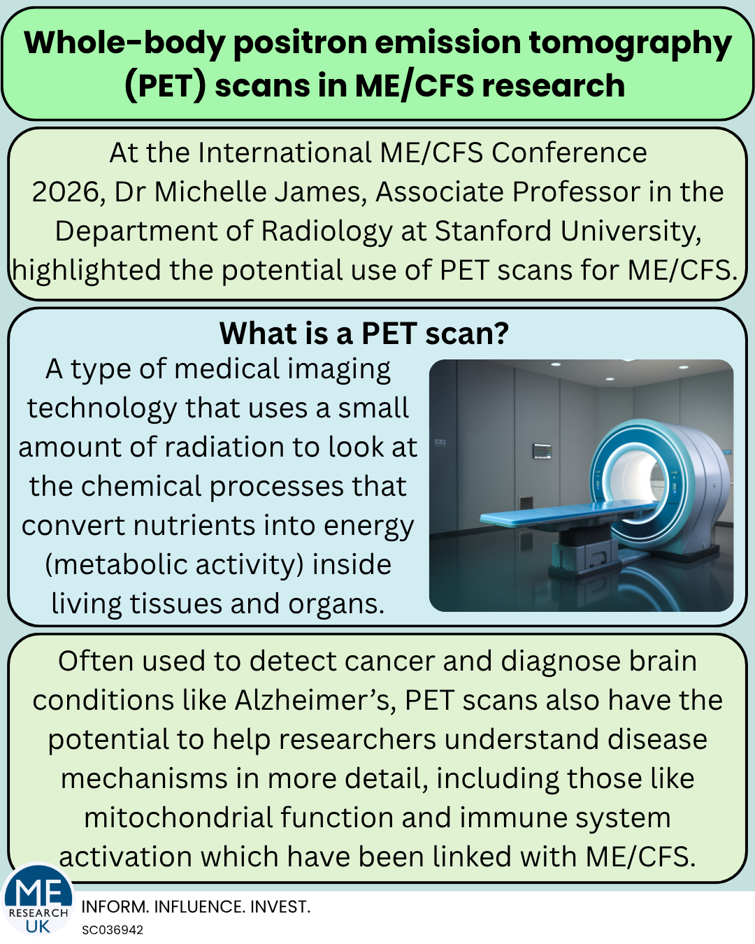
Whole-body positron emission tomography (PET) scans identify differences between people with ME/CFS and healthy controls
Michelle James, PhD spoke about the benefits of the use of whole-body positron emission tomography – or PET – scans for ME/CFS. PET scans use a small amount of radiation to look at metabolic activity of tissues and organs inside the living body. Interestingly, this type of scan has previously been identified as a potential means to detect and quantify the degree of pain associated with disease.
Although not yet published in full, early findings from whole body PET scan research show differences in the signals from muscles between females with ME/CFS and healthy controls, including evidence of ‘coat hanger syndrome’ – a distinctive pattern of pain in the neck and upper back that worsens when upright and often improves when lying down, commonly linked to dysautonomia.
The researchers also identified that there were correlations between signals on the PET scans and the symptoms they measured, including fatigue, pain, and orthostatic intolerance. While this research has only been conducted in female participants so far, the study team also aim to look at males.
Notably in the talk, two biological features detectable in a PET scan – known as PET imaging biomarkers – of interest were highlighted:
- TSPO-PET, which was described as having ‘high potential as a sensor for mitochondrial function and inflammation in ME/CFS’.
- GPR84-PET, which offers a highly promising imaging strategy for the specific detection and tracking of pro-inflammatory microglia and macrophages – both linked with activation of the body’s first, rapid line of defense against pathogens known as the innate immune system.
A video recording of Michelle James talking about PET imaging in ME/CFS for the ME/CFS Collaborative Research Centre at Stanford University may be of interest.